A webinar series to drive demand and adoption of Thermo Fisher Scientific products through education with young scientists in the academia segment

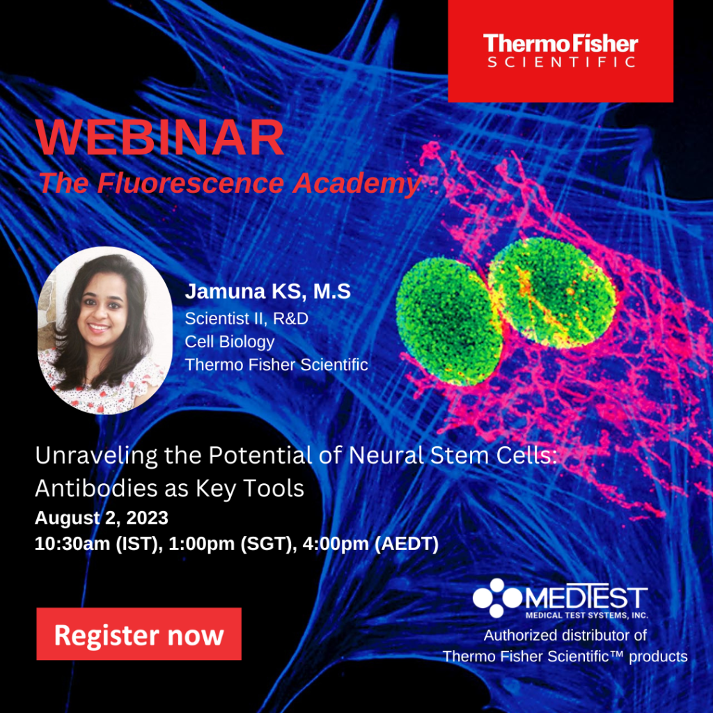
Don’t miss a beat! The second part of the series is here.
Abstract:
Stem cells have the capability to develop into any specialized cell type, which makes them a valuable resource in research and regenerative medicine. Differentiated stem cell models provide insights into proteins involved in disease/developmental processes, which would otherwise be inaccessible for research due to their rare tissue origin and ethical constraints. Embryonic stem cells (ESCs) and induced pluripotent stem cells (iPSCs) differentiated to various lineages serve as powerful tools to study the proteins of interest specific to a cell type. Similarly, neural stem cells (NSCs) have revolutionized the research on neuropsychiatry and neurodegeneration disorders, as they can be differentiated into neuronal subtypes in vitro. In this seminar, we showcased the use of human episomal iPSCs and NSCs to generate physiologically relevant neural models such as retinal ganglion cells, neuroepithelial cells, neural rosettes, and astrocytes. We have also differentiated iPSCs into dopaminergic neurons which help in studying the underlying mechanisms of Parkinson’s disease. These studies are important for deciphering the cellular and molecular mechanisms underlying neurogenesis for optimizing stem cell-based treatment of neurological disorders and injuries.
Learning Objectives
- Outline the overview of pluripotent stem cells and neural stem cells
- Explain the differentiations into committed neural cell types
- Identify the characterization of the differentiated models using antibodies
- Generate disease model for Parkinson’s disease
Webinars will be available for unlimited on-demand viewing after live event.
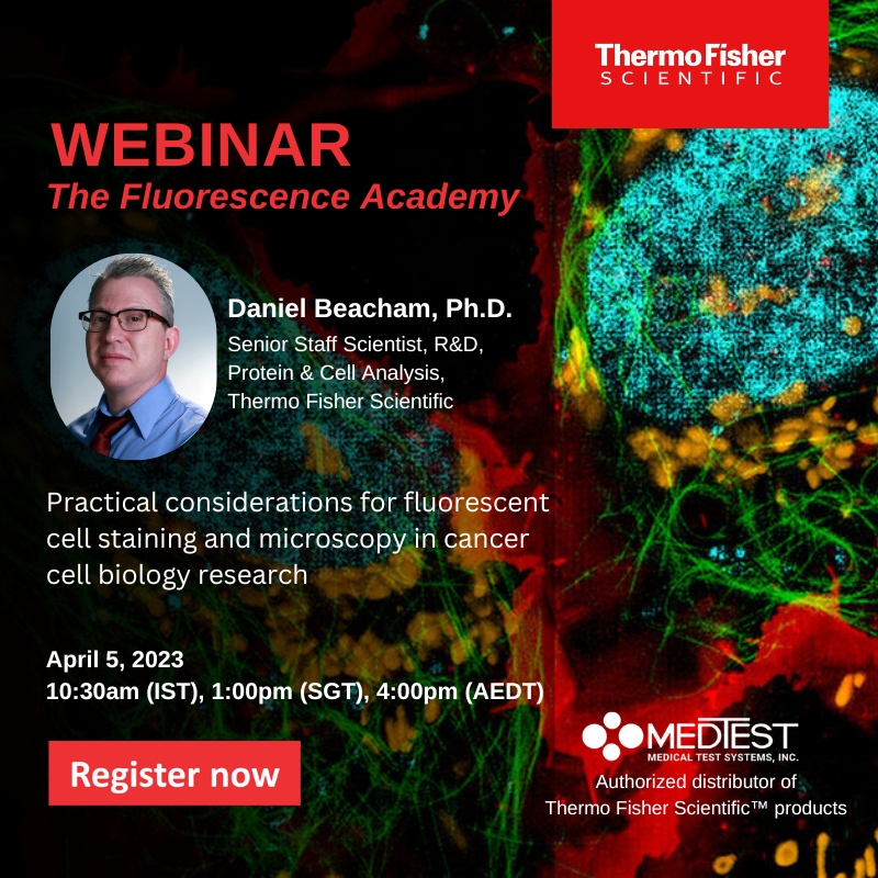
Join the first webinar of the series on April 5th
Abstract:
This seminar provides an overview of fluorescence microscopy in cancer research and other cell-based applications in the biosciences discovery workflow. In comparison with phase contrast and colorimetric cell analysis techniques, fluorescence imaging delivers robust, multi-parametric, and scalable, cell-level analysis of large sample sizes in heterogenous populations with the benefit of spatial and temporal resolution. Following a basic primer on fluorescence imaging and cancer cell biology, a series of existing and newly developed tools for probing cellular structure, cell stress, signaling pathways and function will be reviewed in the context of cancer therapeutics discovery and mechanism of action research. Additional emphasis will be given to the visualization of cell proliferation and cell surface target internalization, including tools for immuno-oncology research applications in both 2D and 3D models.
Learn about the following:
- Basic approaches for fluorescence labeling and microscopy
- Structural and functional tools for cancer cell biology research
- Applications in cancer cell biology research

Can‘t make the live event? Register anyway, and we‘ll send you a link to the recording.
Share this webinar with a colleague »


